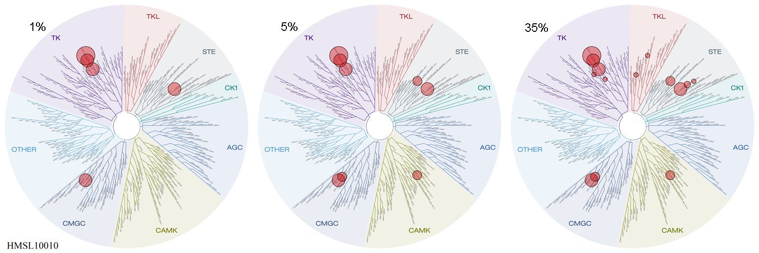
CP724714 KINOMEscan - Dataset (ID:20029)

| HMS Dataset ID: | 20029 |
| Dataset Title: | CP724714 KINOMEscan |
| Project Summary Page(s): | lincs.hms.harvard.edu/kinomescan |
| Screening Lab Investigator: | Qingsong Liu |
| Screening Principal Investigator: | Nathanael Gray |
| Assay Description: | The KINOMEscan assay platform is based on a competition binding assay that is run for a compound of interest against each of a panel of 317 to 456 kinases. The assay has three components: a kinase-tagged phage, a test compound, and an immobilized ligand that the compound competes with to displace the kinase. The amount of kinase bound to the immobilized ligand is determined using quantitative PCR of the DNA tag. Results for each kinase are reported as "Percent of control", where the control is DMSO and where a 100% result means no inhibition of kinase binding to the ligand in the presence of the compound, and where low percent results mean strong inhibition. The KINOMEscan data are presented graphically on TREEspot Kinase Dendrograms (http://www.kinomescan.com/Tools---Resources/Study-Reports---Data-Analysis). For this study, HMS LINCS investigators have graphed results for kinases classified as 35 "percent of control" (in the presence of the compound, the kinase is 35% as active for binding ligand in the presence of DMSO), 5 "percent of control" and 1 "percent of control". |
| Assay Protocol: |
1. T7 kinase-tagged phage strains are grown in parallel in 24-well or 96-well block in a BL21 derived E. coli host for 90 minutes until lysis. 2. Lysates are centrifuged at 6000 g and filtered with a .2 um filter to remove cell debris. 3. Streptavidin-coated magnetic beads are treated with biotinylated kinase ligands for 30 minutes at RT to generate affinity resin. 4. Liganded beads are blocked with excess biotin and washed with blocking buffer (SeaBlock (Pierce) 1% BSA, .05% Tween 20, 1mM DTT) to remove unbound ligand and reduce nonspecific phage binding. 5. Phage lysates, liganded affinity beads, and test compounds are combined in 1X binding buffer (20% SeaBlock, .17 X PBS, .05% Tween 20, 6 mM DTT) in 96-well plates. The final concentration of test compounds is 10 uM. 6. Assay plates are incubated at RT with shaking for 1 hour. 7. Affinity beads are washed 4 X with wash buffer (1X PBS, .05% Tween 20, 1 mM DTT) to remove unbound phage. 8. Beads resuspended after final wash in elution buffer (1X PBS, .05% Tween 20, 2 mM nonbiotinylated affinity ligand) and incubated for 30 minutes at RT. 9. Quantitative PCR is used to measure the amount of phage in each eluate (which is proportional to the amount of kinase bound). Data is presented as % kinase bound by ligand in presence of 10 uM compound compared to DMSO only control reaction. |
| Assay Protocol Reference: |
KINOMEscan website: Overview & Assay Principle Fabian MA, Biggs WH 3rd, Treiber DK, Atteridge CE, Azimioara MD, Benedetti MG, Carter TA, Ciceri P, Edeen PT, Floyd M, Ford JM, Galvin M, Gerlach JL, Grotzfeld RM, Herrgard S, Insko DE, Insko MA, Lai AG, Lelias JM, Mehta SA, Milanov ZV, Velasco AM, Wodicka LM, Patel HK, Zarrinkar PP, Lockhart DJ. A small molecule-kinase interaction map for clinical kinase inhibitors. Nat Biotechnol. 2005 Mar;23(3):329-36. PMID: 15711537 |
| HMS Dataset Type: | KINOMEscan |
| Date Publicly Available: | 2011-07-15 |
| Most Recent Update: | 2015-10-06 |